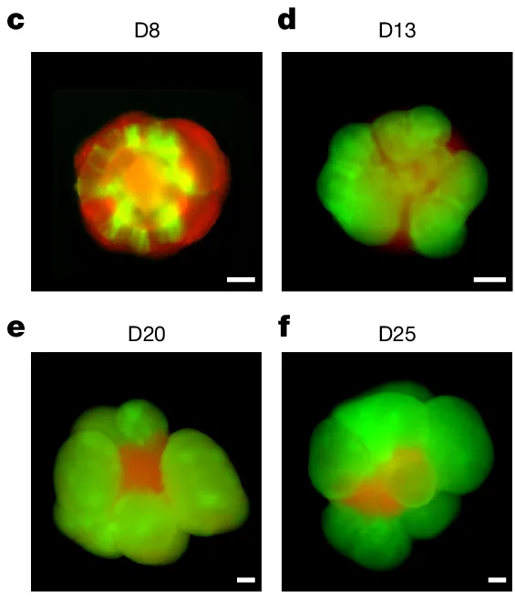
A brain organoid is a small, three-dimensional structure grown from stem cells to model aspects of brain tissue. It can give researchers a way to watch cells divide and specialize in a controlled laboratory setting. Its value comes from the question it helps answer—and from knowing where the model differs from the living tissue it represents.
In a 2026 study, researchers compared a mouse cortical organoid system with development in mice. They found that some cell populations and molecular programs were similar, while the progression of individual stem-cell lineages differed. That result makes the model informative and gives researchers a reason to check its boundaries.
Start with cells that can change
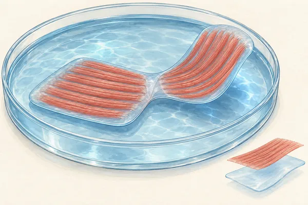
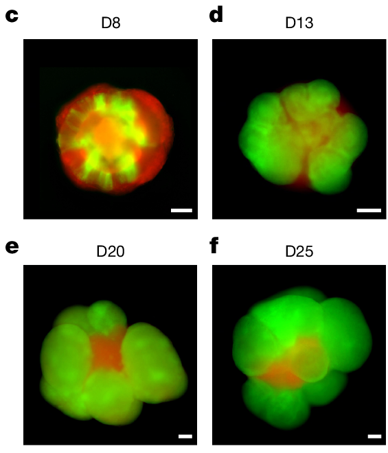
Stem cells can make more cells and produce specialized descendants. Under suitable laboratory conditions, collections of these cells can organize into three-dimensional structures containing cell types and arrangements relevant to a particular organ.
A cortical organoid models aspects of the cerebral cortex, an outer part of the brain. It is not a small complete brain: it does not recreate every region, connection or environmental influence of an animal. Different organoid methods may also create different types of tissue, so a result from one model should be described with its conditions.
For a reader, the useful question is not simply “Does it look brain-like?” Ask instead which cell types, developmental stages and measurements the experiment can study.
Follow a family of cells
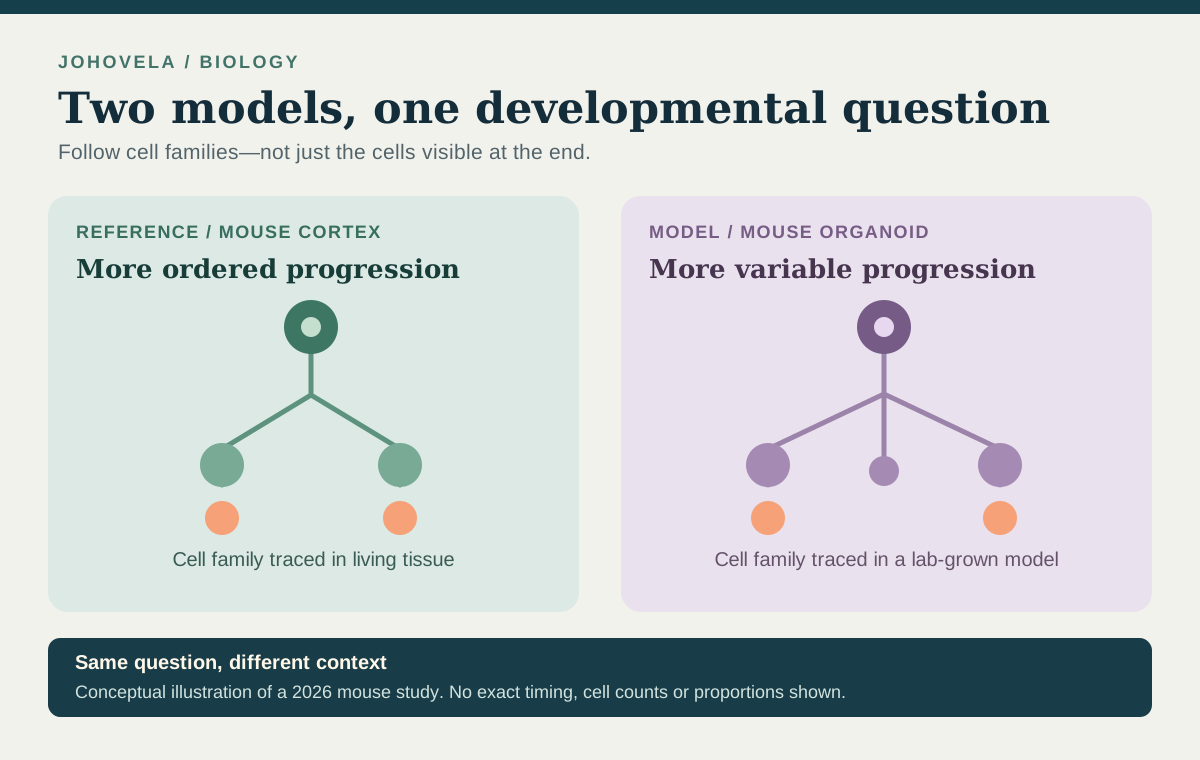
The 2026 team examined radial glial progenitors: cells that divide and give rise to cells in the developing cortex. The researchers adapted a genetic labeling method called mosaic analysis with double markers, or MADM, to trace descendants of individual cells in mouse cortical organoids. Comparing a cell with its descendants is called lineage tracing.
This matters because a snapshot can show that two systems contain similar kinds of cells without showing how each cell family arrived there. Tracing lineages adds information about the divisions and developmental paths that produced those cells.
In the studied organoid system, the researchers observed more variation in the progenitors’ capacity to multiply than in the in-vivo mouse comparison. They also found less variety of cortical projection-neuron types within individual cell families. These are findings about a particular model and its comparison, not a claim that all organoids behave identically.

Similar ingredients can follow a different schedule
The researchers’ institute reports that the organoids and mouse brain shared similar cell populations and molecular programs at the examined stages. Yet the fine timing of progenitor development did not match the same structured progression described for the mouse brain.
An analogy is two kitchens producing a similar meal with different preparation schedules: the ingredients visible at the end do not tell you every step that took place along the way. The analogy helps explain why scientists compare developmental processes as well as final cell types; it is not a measurement of the study.
The researchers suggest that aspects of the stem-cell niche—the surrounding cells, signals and physical environment—may help explain the difference. The study highlights this as an important direction for investigation. It does not identify one proven missing ingredient that accounts for every observed difference.
What the result means for research
Organoids let scientists manipulate and observe developing cells under conditions that can be difficult to examine directly in living tissue. Comparing a model with an appropriate biological reference can show which questions the model is suited to address and where additional evidence is needed.
| Question | Why it matters |
|---|---|
| Which species and tissue? | A mouse cortical model is not automatically a model of every human brain region. |
| What did researchers measure? | Cell types, cell lineages and electrical activity answer different questions. |
| What is the reference? | A comparison with living tissue helps identify similarities and differences. |
| What remains a hypothesis? | A proposed explanation or application still needs its own experiments. |
This is foundational research, not evidence that an organoid can diagnose or treat a person. It also does not establish what subjective experience, if any, an organoid could have. Those questions require separate evidence and careful ethical discussion.
The useful takeaway
An organoid can be valuable without being a complete copy. The mouse study shows why a model’s apparent similarities are a starting point: researchers can investigate developmental differences, test explanations and improve how they interpret future experiments.
Keep the evidence in view.
The highlighted experiment compared a particular mouse cortical organoid system with mouse development. It does not establish the same timing in human organoids or demonstrate a treatment.
Technical sources checked: . Our source check: . This is an AI-assisted editorial explanation, not an independently reproduced experiment.
Sources & further reading
- Nature, August 2026; original researchTemporal uncoupling of radial glia lineage progression in cortical organoids
- Institute of Science and Technology Austria, August 2026Brain and Organoid Side-by-Side